
CELLULAR CHARACTERISTICS UNDER LIGHT MICROSCOPE, ULTRASTRUCTURE
INTRODUCTION
The cell is the functional unit of all living organisms. Simplest organisms i.e. bacteria and algae consist of single cell while more complex organism consist of many cells. Observation of these organelles under light and electron microscope help identify their appearance and their location.
In light microscopy; Conventional light, phase-contrast, differential interference, polarizing, confocal, and fluorescence microscopy are all based on the interaction of light and tissue components. With the light microscope, stained preparations are usually examined by means of light that passes through the specimen. The microscope is composed of mechanical and optical parts. The optical components consist of three systems of lenses: condenser, objective, and eyepiece. The condenser collects and focuses light, producing a cone of light that illuminates the object to be observed. The objective lenses enlarge and project the illuminated image of the object in the direction of the eyepiece. The eyepiece further magnifies this image and projects it onto the viewer's retina, a photographic plate, or (to obtain a digital image) a detector such as a charged coupled device camera. The total magnification is obtained by multiplying the magnifying power of the objective and eyepiece.
A PHASE CONTRAST MICROSCOPE
Some optical arrangements allow the observation of unstained cells and tissue sections. Unstained biological specimens are usually transparent and difficult to view in detail, since all parts of the specimen have almost the same optical density. Phase-contrast microscopy, however, uses a lens system that produces visible images from transparent objects .Phase contrast microscope-uses modified objective lenses and condensers to permit direct examination of living cells without fixation or staining.
Phase contrast microscopes have sub-stage condensers and objectives that convert slight differences in the refractive indices of specimen structure and domains into distinct differences in light intensity
Each organelle and domain in a cell has a unique chemical composition or concentration of chemical constituents which determines its refractive index.
A phase contrast microscope exaggerates these differences so that cell nuclei, cytoplasm ground substance, mitochondria and other cytoplasmic structures appear in contrast to one another.
This type of microscopy does not harm living cells and allows scientists to examine cell behavior under many artificial circumstances
DIFFERENTIAL INTERFERENCE CONTRAST MICROSCOPY
Uses special condensers and objective lenses to transform differences in refractive index into an image that appears to have 3D character.in this type of microscopy, the nucleus and various particulate cytoplasmic inclusions appear in low relief.
FLOURECENCE MICROSCOPY
When certain substances are irradiated by light of a certain wavelength, they emit light with a longer wavelength. This phenomenon is called fluorescence. In fluorescence microscopy, tissue sections are irradiated with either ultraviolet (UV) light or laser, and the emission is in the visible portion of the spectrum. The fluorescent substances appear brilliant or colored on a dark background. Fluorescent compounds that have an affinity for cell macromolecules may be used as fluorescent stains e.g. Acridine orange.
Fluorescence microscopes have a high intensity light source and two filters. The exciter filter, located between the light source and the specimen blocks all light wave lengths except those that excite the flourochromes.
The barrier filter, located between the specimen and the ocular, block all light wavelengths except those emitted by the flourochrome.
Cell structure viewed under microscopes will show same organelles but would appear different depending on the method of microscopy technique used. Different techniques produce relatively different organelle image or structure.
Fluorescence microscopy is most often used to detect specific proteins or other molecules in cells and tissues. A very powerful and widely used technique is to couple fluorescent dyes to antibody molecules, which then serve as highly specific and versatile staining reagents that bind selectively to the particular macromolecules they recognize in cells or in the extracellular matrix.
POLARIZING MICROSCOPY
Polarizing microscopy allows structures made of highly organized molecules to be recognized. When normal light passes through a polarizing filter (such as a Polaroid), it excites vibration in only one direction. If a second filter is placed in the microscope above the first one, with its main axis perpendicular to the first filter, no light passes through. If, however, tissue structures containing oriented molecules (such as cellulose, collagen, microtubules, and microfilaments) are located between the two polarizing filters, their repetitive, oriented molecular structure rotates the axis of the light emerging from the polarizer. Consequently, they appear as bright structures against a dark background
CONFOCAL MICROSCOPY
The depth of focus in the regular light microscope is relatively long, especially when small magnification objectives are used. This means that a rather wide extent of the specimen is seen in focus, simultaneously causing superimposition of the image of a three-dimensional object. With confocal microscopy, on the other hand, only a very thin plane of the specimen is seen in focus at one time. There are two principles on which this is based: (1) the specimen is illuminated by a very small beam of light (whereas in the common light microscope, a large beam of light floods the specimen) and (2) the image collected from the specimen must pass through a small pinhole. The result is that only the image originating from the focused plane reaches the detector whereas the images in front of and behind this plane are blocked


CELLULAR COMPONENTS:
1.The nucleus
Is seen as a dense body in most cells and is the largest organelle in the cells.
Cell nucleus under light microscope
There is a great variety in the size of the nucleus, its shape, the densities of staining of chromatin, nucleolus and in the position of the sex chromatin.
2.Cytoplasm
Is called the soluble phase of the cell, consisting of mostly water, dissolved solutes and larger molecules in suspension tending to link repetitively with covalent bonds giving the cytoplasm a dense, viscous, colloidal sol or gel consistency.
3.Endoplasmic Reticulum
Are cytoplasmic membrane system of many parallel membranes and tubules in communication with each other and lead towards the Golgi apparatus. Are of two types:
Rough endoplasmic reticulum-has fine granules (ribosomes), in clusters studding the outer surface of the parallel membrane and are related to protein synthesis.
Smooth endoplasmic reticulum- lack ribosomes and is more tubular. Is associated with protein synthesis.
4.Golgi body/ complex/ Apparatus
Usually takes one place near to the nucleus and often in a specific place. Consist of a complex of stacked smooth cisternae- enclosing lamellae, tubules and vesicles of various sizes. In light microscope, after special staining (silver), the Golgi apparatus may be seen as a tangled network. Is involved in concentration and preparation for storage of proteins and completing the synthesis of complex sugars
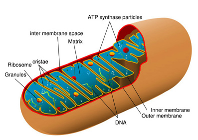
5.Mitochondria
In light microscope can be stained with special methods and appear as coloured rods or granules are hollow bodies enclosed in two unit membranes. Are rich in the enzymes associated with the storage and release of energy and with respiration and fatty acid metabolism may also store calcium.
6.Filaments
Are fine threads visible in electron microscope but may be exaggerated into thicker myofibrils visible in light microscope. Are very important contractile elements in all cells as they furnish a flexible skeleton articulated by the cell itself.
7.Centrosome/ division body
A body which lies alongside the nucleus at the cell center or cytocentum and is just visible as dots in light microscope
8.Lysosomes
Are round, single membrane-limited, darkly staining bodies without cristae and containing another class of enzyme- hydrolytic or digestive.
9.Cell inclusions
Non-living, non-participating, poorly structured cell elements, very rarely seen in an intra-nuclear position, usually cytoplasmic e.g.
a)Fat droplets- appearing as vacuoles in ordinary light microscope preparation.
b)Lamella bodies- contain lipids to be secreted.
c)Glycogen granules
d)Secretion granules- e.g. zymogen in pancreatic cells.
e)Pigments- melanin(skin cells), lipofuscin (old neurons), carbon(exogenous)
f)Crystals-testicular interstitial cells.
g)Bacterial and viral inclusion bodies(pathological)
CONCLUSION
The light microscope, so called because it employs visible light to detect small objects, is probably the most well-known and well-used research tool in biology. The main groups of techniques are small chemical staining of cellular structures, for example DAPI to label DNA, use of antibodies conjugated to fluorescent reporters and fluorescent proteins, such as green fluorescent proteins. These techniques use these different fluorophores for analysis of cell structure at a molecular level in both live and fixed samples.
You can add more text, to the conclusion as this is a plain and short conclusion.
.
Informative post about Cellular Characteristics under light microscope. While purchasing a microscope a you should keep in mind their hobbies and the reason for which he is buying.
ReplyDeleteSimilar to the cue ball that is shot in pools; this game
ReplyDeleteinvolves coins and strikers that are essential to play carrom game.
Age Level - A good factor to write about is the appropriate age level for
the video game that you are reviewing. But what happens when a player runs out of race tokens.
Also visit my blog ... http://www.sandbridgepier.com/sbforum/profile.php?id=27254
There are also significant role that your cake can play in your wedding day.
ReplyDeleteLater, during the Middle Ages, many guests would bring small cakes to the wedding celebration.
Since the machines can also cut logos, letters and signs, the cake creator can
later also decorate the.
Have a look at my web blog ... cake art salisbury md
clients, Rachel, who got back together with her ex. The only downside of it in a lot
ReplyDeleteof people's eyes is the fact that there is no multiplayer content. Are you a Kansas City Chiefs or Pittsburgh Steelers fan.
Feel free to visit my web-site; Continued :: ::
If some one wishes to be updated with most up-to-date technologies
ReplyDeletethen he must be pay a visit this web page and be up to date every day.
Here is my blog post Dr Dre Beats Pro
Great Article it its really informative and innovative keep us posted with new updates. its was really valuable. thanks a lot.
ReplyDeleteChandeliers