Showing posts with label Human Physiology. Show all posts

Wednesday, May 30, 2012

Resting Membrane Potential of Nerves


The resting membrane potential of large nerve fibers when not transmitting nerve signals is about –90 millivolts. That is, the potential inside the fiber is 90 millivolts more negative than the potential in the extracellular fluid on the outside of the fiber. In the next few paragraphs, we explain all the factors that determine the level of this resting potential, but before doing so, we must describe the transport properties of the resting nerve membrane for sodium and potassium.
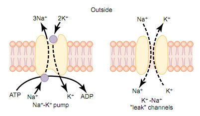
Active Transport of Sodium and Potassium Ions through the Membrane—the Sodium-Potassium (Na+ -K+) Pump. First, let us recall that all cell membranes of the body have a powerful Na+ -K+ that continually pumps sodium ions to the outside of the cell and potassium ions to the inside, as illustrated on the left-hand side in. Further, note that this is an electrogenic pump because more positive charges are pumped to the outside than to the inside (three Na+ ions to the outside for each two K+ ions to the inside), leaving a net deficit of positive ions on the inside; this causes a negative potential inside the cell membrane The Na+ -K+ also causes large concentration gradients for sodium and potassium across the resting nerve membrane Leakage of Potassium and Sodium Through the Nerve Membrane. The right side of Figure below shows a channel protein in the nerve membrane through which potassium and sodium ions can leak, called a potassium-sodium (K+ -Na+) “leak” channel. The emphasis is on potassium leakage because, on average, the channels are far more permeable to potassium than to sodium, normally about 100 times as permeable. As discussed later, this differential in permeability is exceedingly important in determining the level of the normal resting membrane potential.
Origin of the Normal Resting Membrane Potential
The figure shows the important factors in the establishment of the normal resting membrane potential of–90 millivolts. They are as follows.
1.      Contribution of the Potassium Diffusion Potential.
In the figure below1A, we make the assumption that the only movement of ions through the membrane is diffusion of potassium ions, as demonstrated by the open channels between the potassium symbols (K+) inside and outside the membrane. Because of the high ratio of potassium ions inside to outside, 35:1, the Nernst potential corresponding to this ratio is –94 millivolts because the logarithm of 35 is 1.54, and these times –61millivolts is –94 millivolts. Therefore, if potassium ions were the only factor causing the resting potential, the resting potential inside the fiber would be equal to –94 millivolts, as shown in the figure.


2.      Contribution of Sodium Diffusion through the Nerve Membrane.
            The figure shows the addition of slight permeability of the nerve membrane to sodium ions, caused by the minute diffusion of sodium ions through the K+ -Na leak channels. The ratio of sodium ions from inside to outside the membrane is 0.1, and this gives a calculated Nernst potential for the inside of the membrane of +61 millivolts. But also shown in Figure 1B is the Nernst potential for potassium diffusion of –94 millivolts. How do these interact with each other, and what will be the summated potential? This can be answered by using the Goldman equation described previously. Intuitively, one can see that if the membrane is highly permeable to potassium but only slightly permeable to sodium, it is logical that the diffusion of potassium con tributes far more to the membrane potential than does the diffusion of sodium. In the normal nerve fiber, the permeability of the membrane to potassium is about 100 times as great as its permeability to sodium. Using this value in the Goldman equation gives a potential inside the membrane of –86 millivolts, which is near the potassium potential shown in the figure.

3.      Contribution of the Na -K Pump.
In Figure 1C, the Na+ -K+ pump is shown to provide an additional contribution to the resting potential. In this figure, there is continuous pumping of three sodium ions to the outside for each two potassium ions pumped to the inside of the membrane. The fact that more sodium ions are being pumped to the outside than potassium to the inside causes continual loss of positive charges from inside the membrane; this creates an additional degree of negativity (about –4 millivolts additional) on the inside beyond that which can be accounted for by diffusion alone. Therefore, as shown in Figure 1C, the net membrane potential with all these factors operative at the same time is about –90 millivolts.
In summary, the diffusion potentials alone caused by potassium and sodium diffusion would give a membrane potential of about –86 millivolts, almost all of this being determined by potassium diffusion. Then, an additional  –4 millivolts is contributed to the membrane potential by the continuously acting electrogenic Na+ -K+ pump, giving a net membrane potential of –90 millivolts.

CELL DIFFERENTIATION


A special characteristic of cell growth and cell division is cell differentiation, which refers to changes in physical and functional properties of cells as they proliferate in the embryo to form the different bodily structures and organs. The description of an especially interesting experiment that helps explain these processes follows.
When the nucleus from an intestinal mucosal cell of a frog is surgically implanted into a frog ovum from which the original ovum nucleus was removed, the result is often the formation of a normal frog. This demonstrates that even the intestinal mucosal cell, which is a well-differentiated cell, carries all the necessary genetic information for development of all structures required in the frog’s body.
Therefore, it has become clear that differentiation results not from loss of genes but from selective repression of different genetic operons. In fact, electron micrographs suggest that some segments of DNA helixes wound around histone cores become so condensed that they no longer uncoil to form RNA molecules. One explanation for this is as follows: It has been supposed that the cellular genome begins at a certain stage of cell differentiation to produce a regulatory protein that forever after represses a select group of genes. Therefore, the repressed genes never function again. Regardless of the mechanism, mature human cells produce a maximum of about 8000 to 10,000 proteins rather than the potential 30,000 or more if all genes were active.
Embryological experiments show that certain cells in an embryo control differentiation of adjacent cells. For instance, the primordial chorda-mesoderm is called the primary organizer of the embryo because it forms a focus around which the rest of the embryo develops.
It differentiates into a mesodermal axis that contains segmentally arranged somite’s and, as a result of inductions in the surrounding tissues, causes formation of essentially all the organs of the body.
Another instance of induction occurs when the developing eye vesicles come in contact with the ectoderm of the head and cause the ectoderm to thicken into a lens plate those folds inward to form the lens of the eye. Therefore, a large share of the embryo develops as a result of such inductions, one part of the body affecting another part, and this part affecting still other parts.
Thus, although our understanding of cell differentiation is still hazy, we know many control mechanisms by which differentiation could occur.

APOPTOSIS—PROGRAMMED CELL DEATH


The 100 trillion cells of the body are members of a highly organized community in which the total number of cells is regulated not only by controlling the rate of cell division but also by controlling the rate of cell death. When cells are no longer needed or become a threat to the organism, they undergo a suicidal programmed cell death, or apoptosis. This process involves a specific proteolytic cascade that causes the cell to shrink and condense, to disassemble its cytoskeleton, and to alter its cell surface so that a neighboring phagocytic cell, such as a macrophage, can attach to the cell membrane and digest the cell.
In contrast to programmed death, cells that die as a result of an acute injury usually swell and burst due to loss of cell membrane integrity, a process called cell necrosis. Necrotic cells may spill their contents, causing inflammation and injury to neighboring cells. Apoptosis, however, is an orderly cell death that results in disassembly and phagocytosis of the cell before any leakage of its contents occurs, and neighboring cells usually remain healthy.
Apoptosis is initiated by activation of a family of proteases called caspases. These are enzymes that are synthesized and stored in the cell as inactive procaspases. The mechanisms of activation of caspases are complex, but once activated; the enzymes cleave and activate other procaspases, triggering a cascade that rapidly breaks down proteins within the cell. The cell thus dismantles itself, and its remains are rapidly digested by neighboring phagocytic cells.

A tremendous amount of apoptosis occurs in tissues that are being remodeled during development. Even in adult humans, billions of cells die each hour in tissues such as the intestine and bone marrow and are replaced by new cells. Programmed cell death, however, is precisely balanced with the formation of new cells in healthy adults. Otherwise, the body’s tissues would shrink or grow excessively. Recent studies suggest that abnormalities of apoptosis may play a key role in neurodegenerative diseases such as Alzheimer’s disease, as well as in cancer and auto-immune disorders. Some drugs that have been used successfully for chemotherapy appear to induce apoptosis in cancer cells.